Hemodynamic lag assessment is proving to be an informative tool for looking at various cerebral pathologies. In this incredible video, a peri-operative recording of an STA-MCA bypass performed on a pediatric moyamoya patient using ICG-enhanced fluorescence is shown.
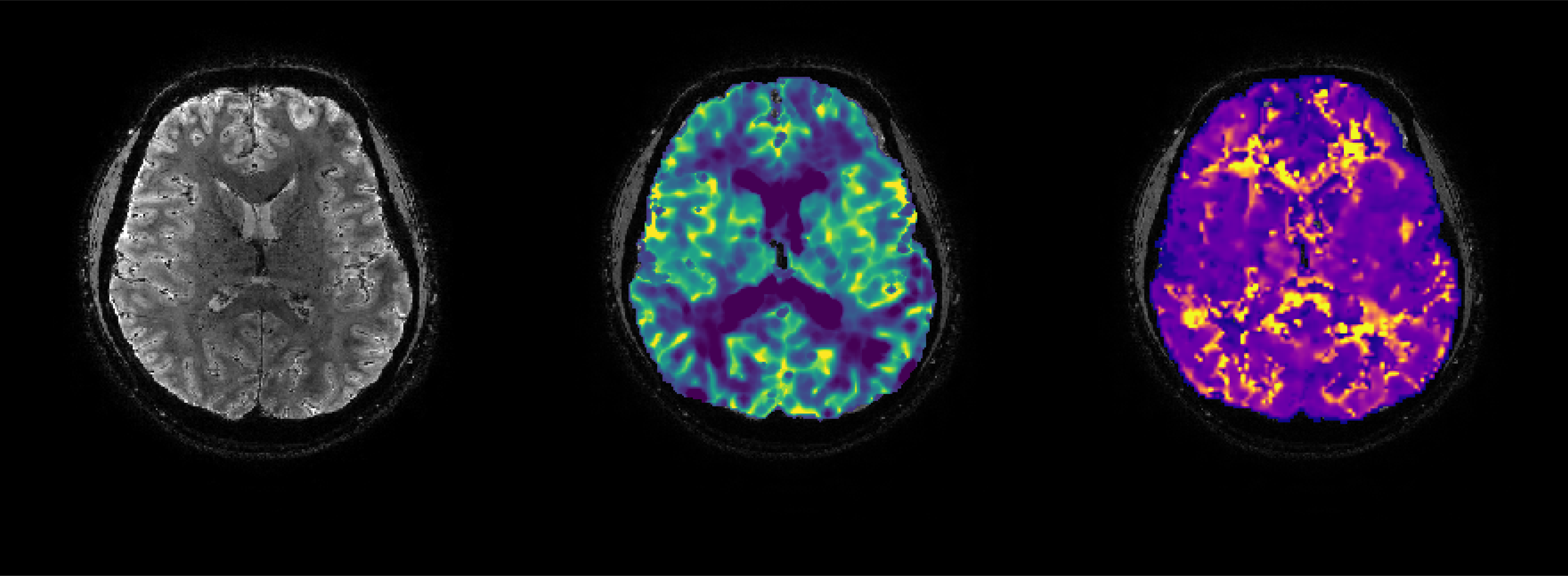
Pre-operatively the CBF and CVR of the patient was measured using BOLD and ASL in combination with acetazolamide. This showed a steal phenomenon so an operation was planned. During this operation the main branch of the external carotid artery; the superficial temporal artery was connected to a distal middle cerebral artery, forming a direct extra-cranial to intracranial bypass. To test the patency of the bypass, ICG-enhanced fluorescence was used to visualize the flow from the bypass to the brain. When you look closely you can see the blood coming from the bypass entering the brain before it arrives from
the internal carotid; highlighting the delayed transit time often seen in moyamoya vasculopathy. With the extra blood flow of the bypass, the aim is to re-vascularize the cortex and restore the cerebrovascular reserve, to ultimately reduce the number of TIA’s and risk of infarction. Note the time delay between blood arrival through the bypass and the eventual drainage through the large pial veins covering on the surface of the cortex. See the tutorials for analysis pipelines designed to visualize hemodynamic delay in patients such as these.
*video provided by Pieter Deckers

